HPI
30 yo Vietnamese female presented to cornea clinic for evaluation of red eyes. She was having some eye redness and clouding of her vision recurrently over the past 4 years when she was in Vietnam. She had some “laser” procedure done at that time and then had been advised to use PredForte as needed which the patient did until she ran out 5 days prior to presentation. Current flare started two weeks ago. Patient does not speak English well.
She states she has only been symptomatic in her right eye, although both do get red. She has never used PredForte in her left eye. She denies using any oral prednisone or other immunosuppressant medication, also denies periocular injections of medications.
She denies any family history of uveitis
No current systemic medications or reported drug allergies
Currently lives in Pittsburgh with her boyfriend and is employed doing nails.
ROS
Patient does complain of whitening her hair around the crown recently and has been dying that area, however denies any skin rash or vitiligo, denies any tinnitus.
Denies TB exposure, although patient is not really sure what TB is. Denies oral ulcerations.
Denies other systemic complaints including dizziness, tinnitus, meningismus, palpitations, shortness of breath, diarrhea, nausea, dysuria, claudication.
|
||||||||||||||||||||||||||||||||||||||||||||||||||||||||||||||||||||||||||||||||||
|
Main Ophthalmology Exam
|
|||
|
|
External Exam
|
||
|
|
Right
|
Left
|
|
|
|
External
|
Normal
|
Normal
|
|
|
Slit Lamp Exam
|
||
|
|
Right
|
Left
|
|
|
|
Lids/Lashes
|
Normal
|
Normal
|
|
|
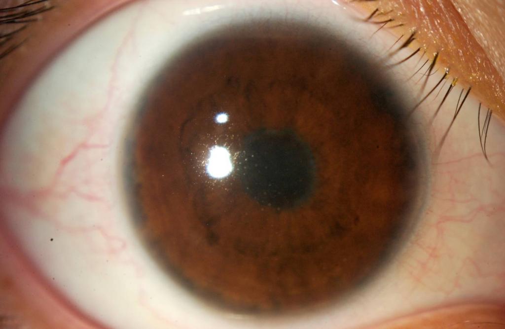
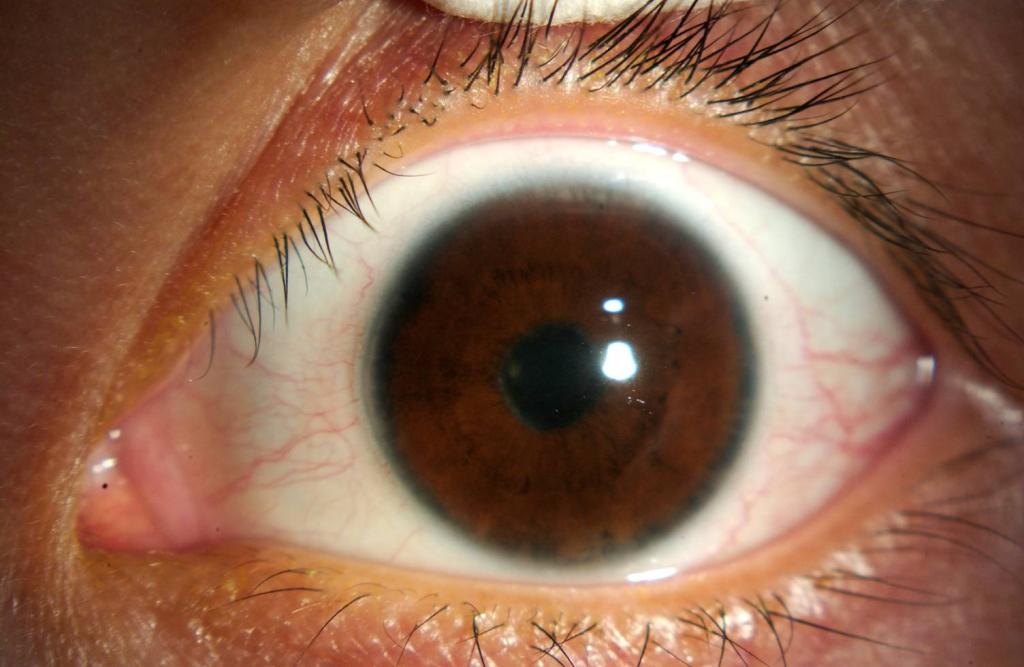
Conjunctiva/Sclera
|
3+ Injection
|
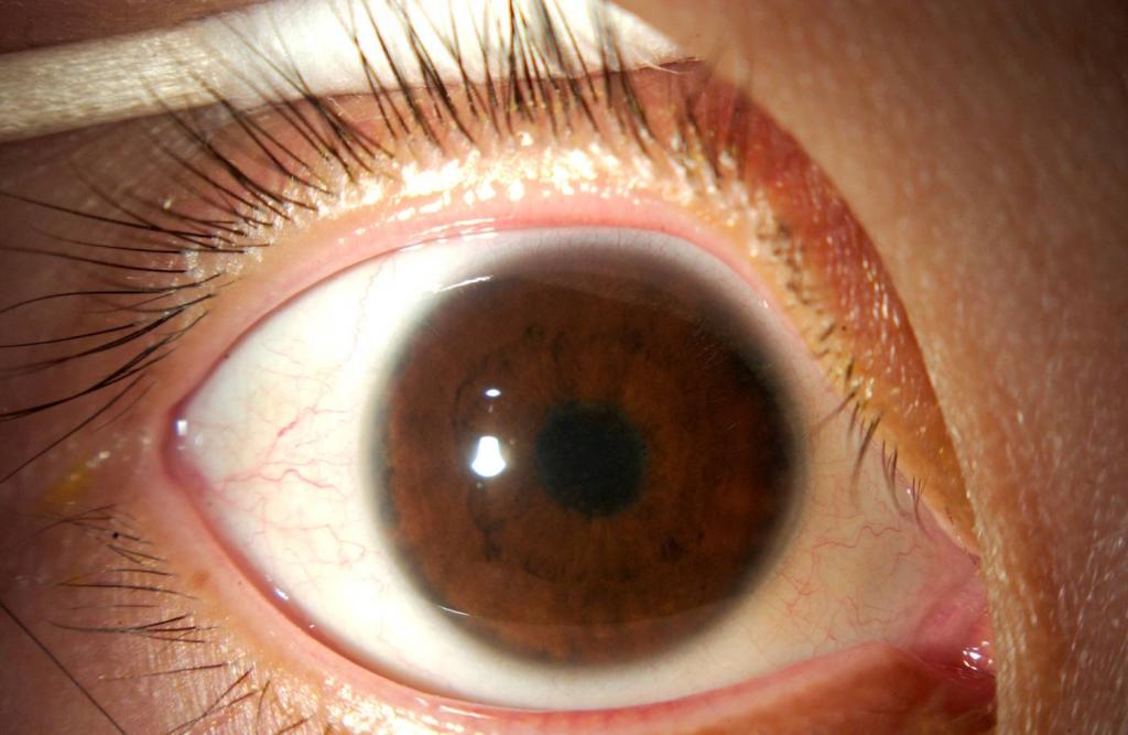
1+ Injection
|
|
|
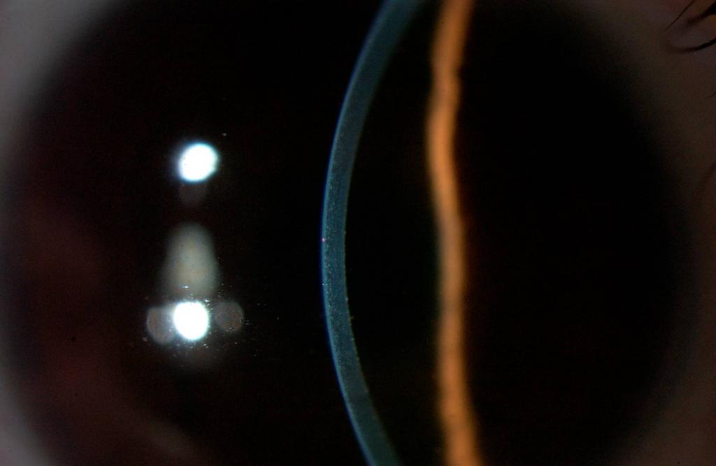
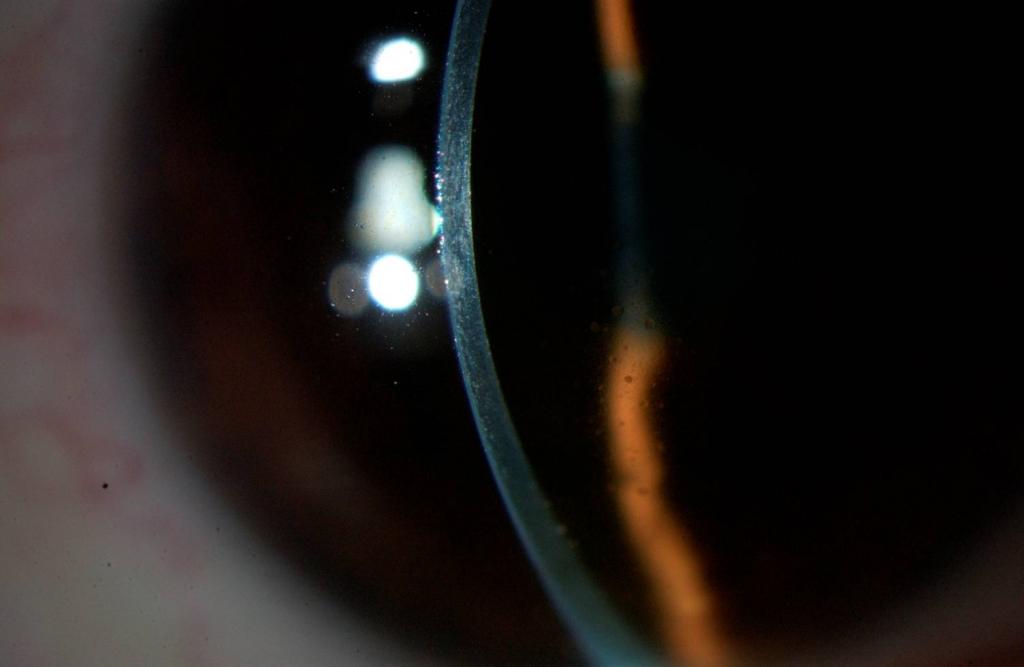
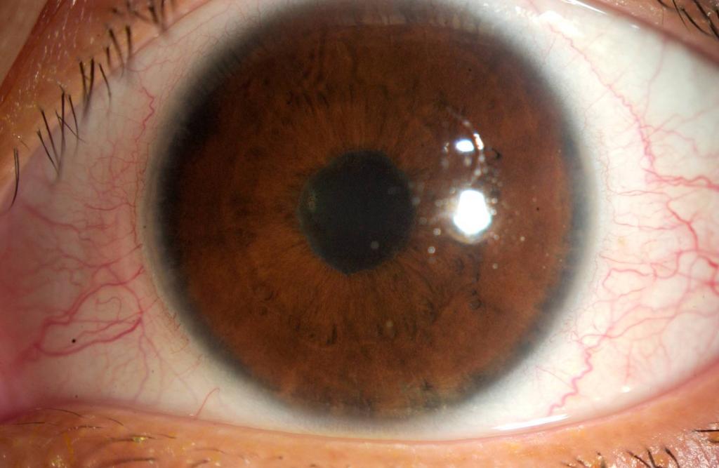
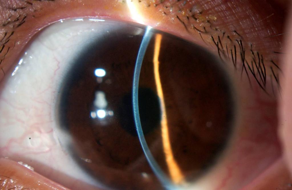
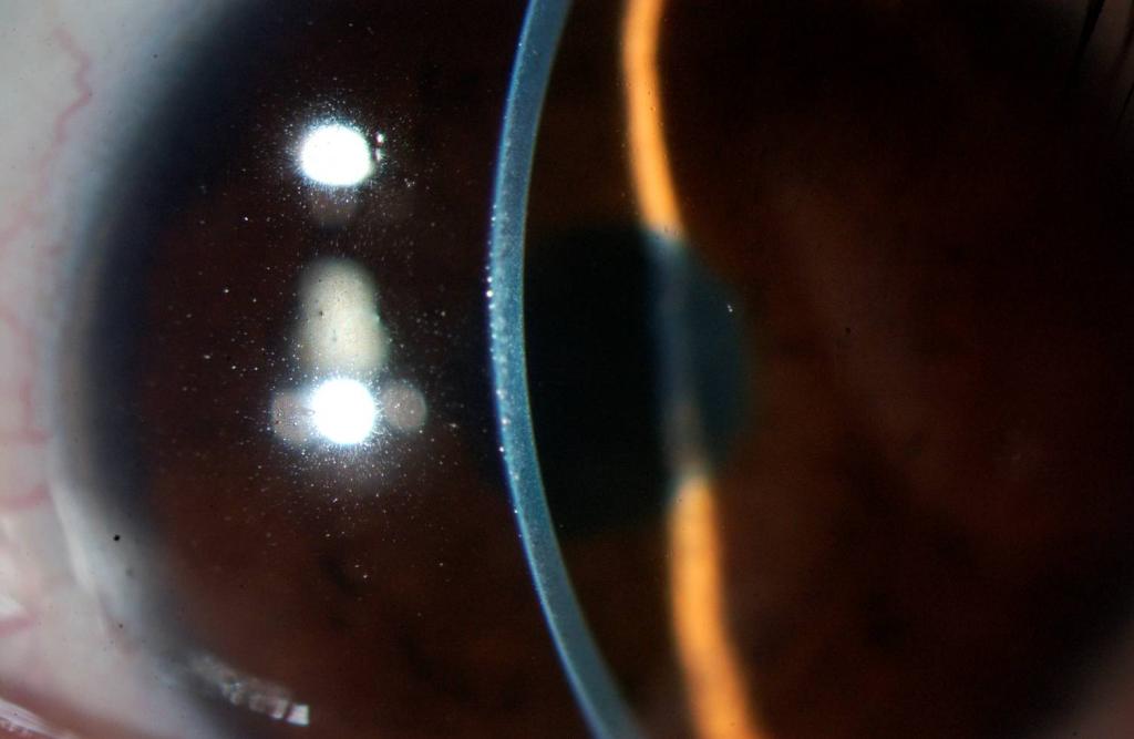
Cornea
|
2+ Keratic precipitates (granulomatous)
|
Trace Keratic precipitates
|
|
|
Anterior Chamber
|
3+ Cell /3+flare
|
Trace flare
|
|
|
Iris
|
Secluded pupil, peripheral atrophy (laser gonioplasty?), PI patent
|
Secluded pupil, peripheral atrophy (laser gonioplasty?), PI patent
|
|
|
Lens
|
Nuclear Sclerosis
|
Nuclear Sclerosis
|
|
|
Vitreous
|
limited view without obvious cell
|
limited view no cell
|
|
|
Fundus Exam
|
||
|
|
Right
|
Left
|
|
|
|
Disc
|
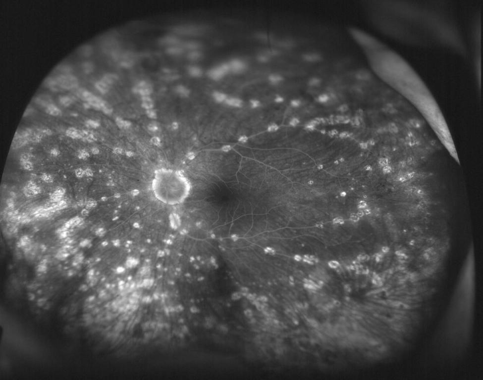
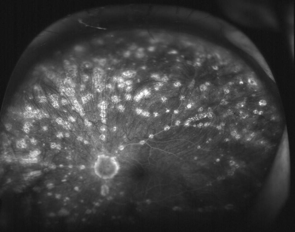
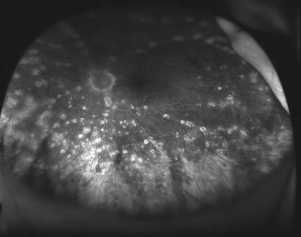
Peripapillary atrophy 360 degrees C/D 0.1
|
Peripapillary atrophy 360 degrees C/D 0.1
|
|
|
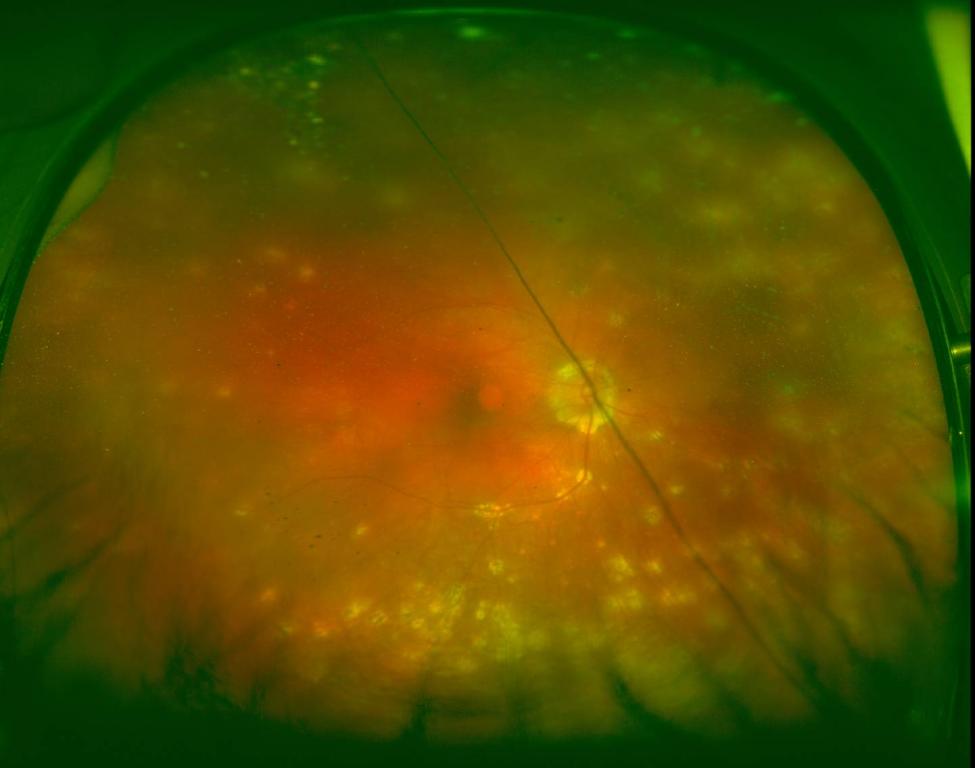
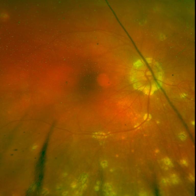
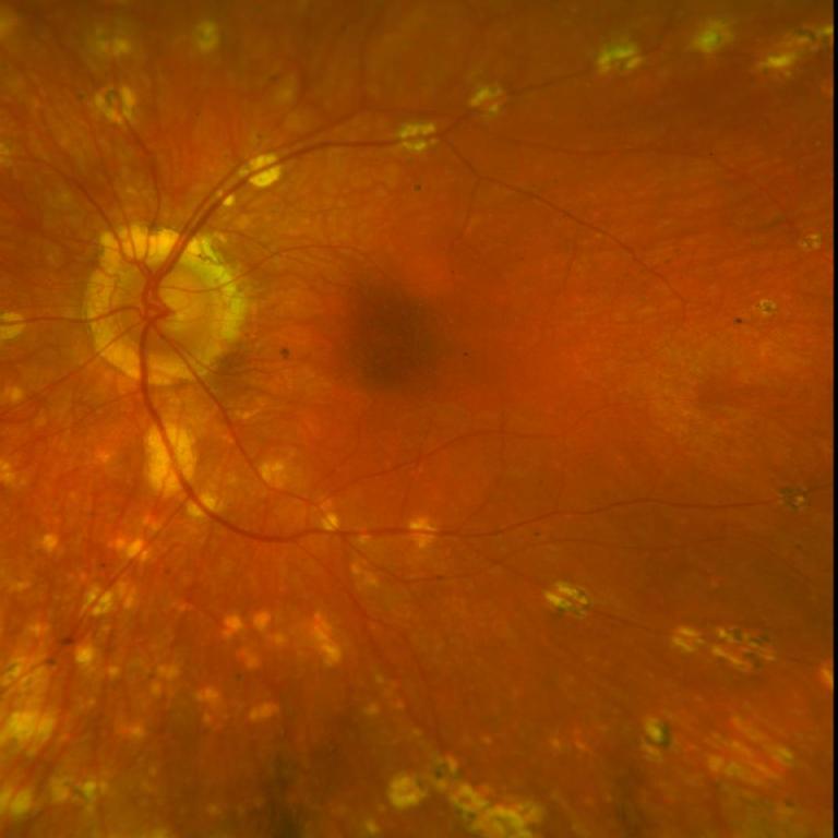
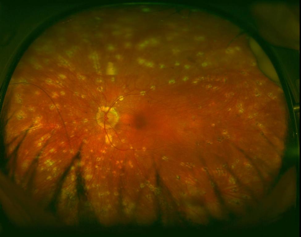
Macula
|
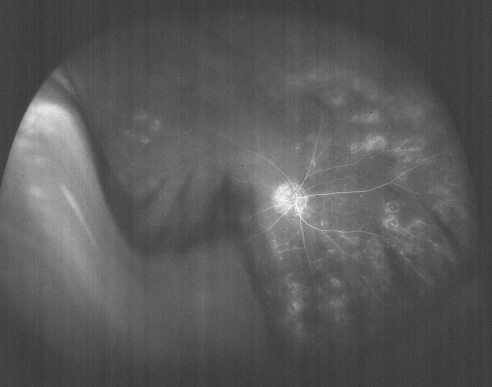
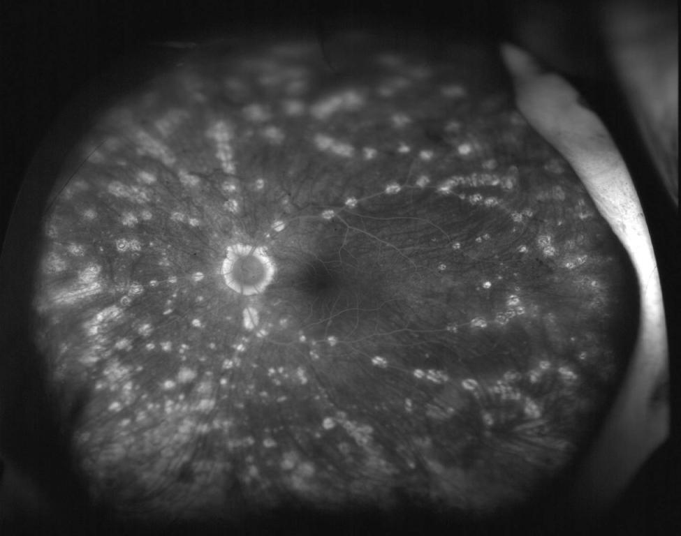
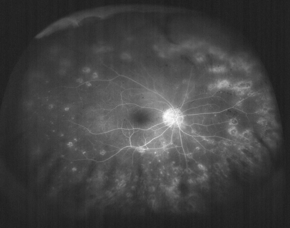
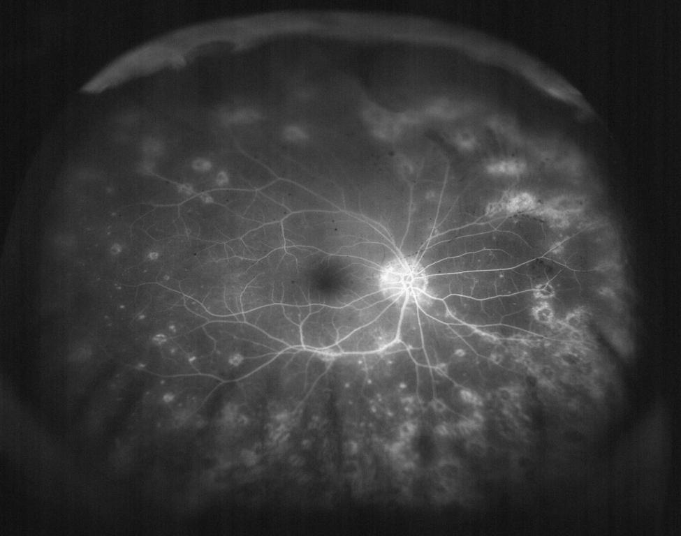
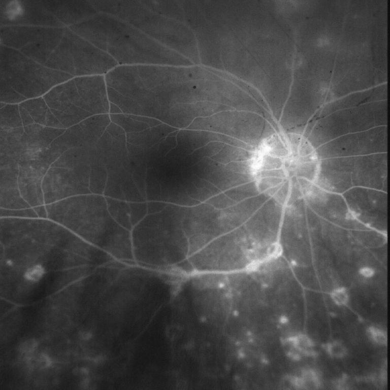
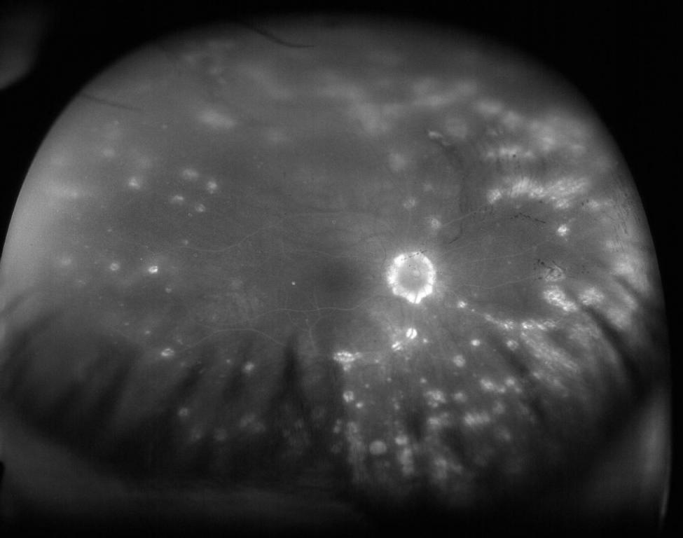
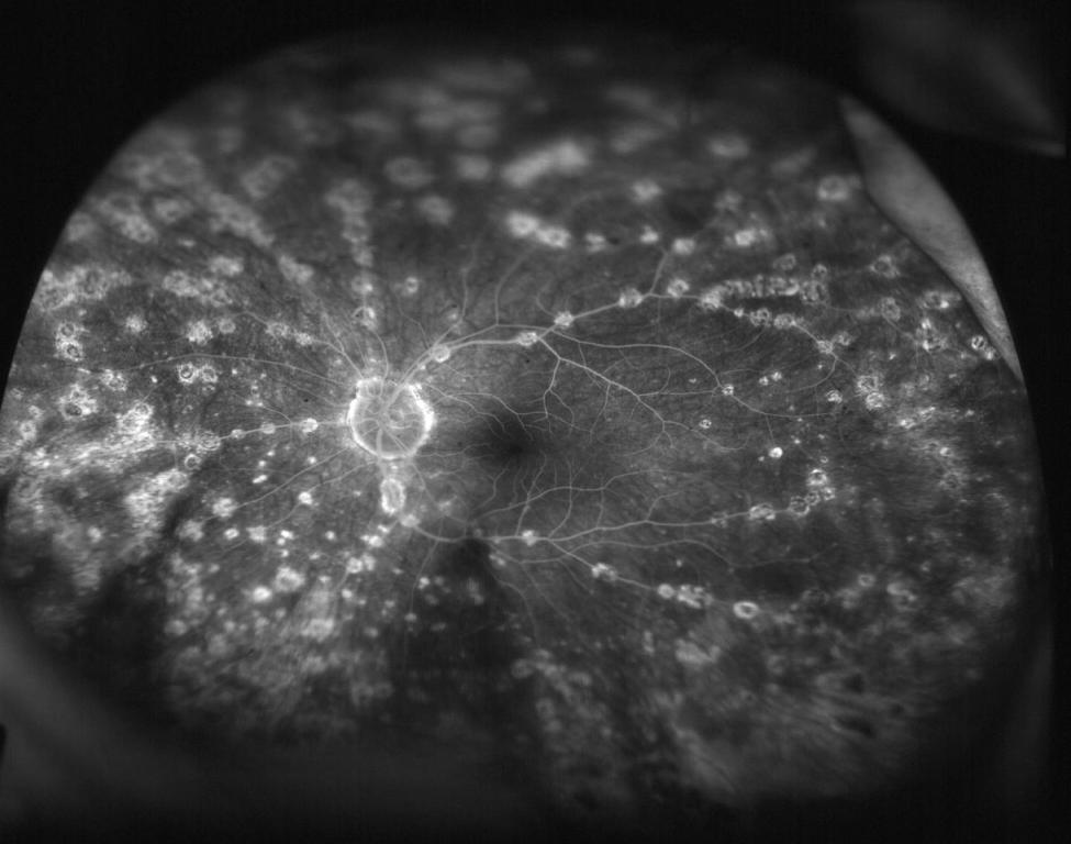
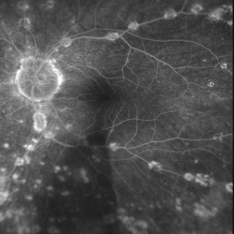
chorioretinal punched out lesions tracking along vessels and throughout periphery but sparing macula
|
chorioretinal punched out lesions tracking along vessels and throughout periphery but sparing macula symmetrical to contralateral eye
|
|
|
Vessels
|
no active sheathing
|
no active sheathing
|
|
|
Periphery
|
limited by secluded pupil
multiple punched out lesions with pigmentary chagnes, no active infiltrates; blonde fundus
|
limited by secluded pupil
mutliple punched out lesions with pigmentary changes, no active infiltrates; blond fundus
|
Laboratory/Radiographic Evaluation:
Chest XRay - Normal
CBC, Basic Metabolic Panel WNL
RPR, Lyme ELISA Negative
ANA <1/80; ESR 8
HLA-DR4 Positive -- HLA DRB1*04, HLA-DRB1*12
HLA-B27 Negative
ACE, ANCA Pending